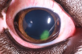
Non-healing corneal ulcers
In an uncomplicated, superficial ulcer, the epithelium (surface layer) migrates across the exposed stroma (middle layer) and creates attachments as it grows. Non-healing corneal ulcers, also known as indolent ulcers, spontaneous chronic corneal epithelial defects (SCCEDs), or boxer ulcers, occur spontaneously with no known trauma, and are often due to the presence of an acellular hyalinized membrane on the surface of the stroma (middle layer of the cornea). This membrane interferes with adhesion formation between the migrating epithelial cells and underlying stroma, thus preventing healing. Most indolent ulcers require some procedural intervention to remove this membrane and stimulate healing.
 In general, there are three procedures, which can be used in various combinations in the clinic without sedation or general anesthesia. These include a cotton-tipped applicator debridement (30-50% success of healing); diamond burr debridement (75% success of healing), and a grid keratotomy (combined with the diamond burr, 90-95% success of healing). In rare cases, the area of indolent ulcers require surgical removal (superficial keratectomy), although most cases are successful with one of the less invasive procedures.
After the procedure, we often place a bandage contact lens to cover the exposed nerve endings to improve comfort. Post-procedural care includes topical antibiotics, systemic pain medications with or without anti-inflammatories, and an E-collar (cone). Some patients are additionally sent home with supplemental lubrication. It is critical that an E-collar be worn at all times until recheck, as any rubbing or pawing can disrupt the healing epithelium. Patients are rechecked in 10-14 days, and contact lenses are removed if still present.
Infected corneal ulceration
The cornea is the transparent, domed portion of the eye that acts to protect and maintain the ocular contents while allowing light to enter the eye for vision. The cornea is made up of four layers: the outer epithelial layer, middle stromal layer, and deep Descemet’s membrane and endothelial layers.
An infected corneal ulceration affects the mid to deeper layers of the cornea. Typically, this process starts with a specific underlying cause such as trauma, dry eye, or eyelid abnormalities leading to an abrasion on the surface of the eye. Once the surface layer of the cornea is disrupted it allows for bacterial access to the deeper layers. This bacterial infection can cause significant breakdown of the cornea and potential compromise and loss of the eye. Thus, aggressive treatment is needed. Cytology and culture of the ulceration are recommended before instituting topical antibiotic therapy. Depending on the severity of your pet’s ulceration either medical and/or surgical management will be recommended.
Medical management
Generally, if your pet’s ulceration is affecting less than 50% of the corneal depth, medical management is recommended. Once corneal cytology and culture have been performed, the eye is treated with broad-spectrum topical antibiotics. Topical agents such as serum or EDTA will be recommended to prevent further breakdown of the cornea (also known as corneal melting). Topical atropine is recommended to decrease pain and dilate the pupil. Additional medications may be added as necessary. Medications are typically started at a frequency of every two hours with five minutes between each drop. Regular rechecks are required, with the first recheck within 24-72 hours of diagnosis.
Surgical management
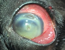 Deep corneal ulceration created a weak spot that is at risk for catastrophic corneal perforation. If your pet’s ulceration is affecting more than 50% of the corneal depth, surgical management in the form of a conjunctiva graft is recommended. This is a procedure performed under general anesthesia in which a piece of the pink tissue surrounding the clear cornea is sutured over the corneal ulceration (see photo). Sometimes your dog may require topical medical treatment for 24-48 hours prior to the procedure to better ensure success. There is an 85-90% chance of success (healing of the eye) with this procedure, but complications, such as graft failure, are possible. Topical medications four times daily are recommended following surgery, along with one- and three-week post-operative rechecks.